Overview of Syllabus Under Master of Science in Radio and Imaging Technology Year 1

Updated Post: 04 May 2024
Share Post:
![]()
![]()
![]()
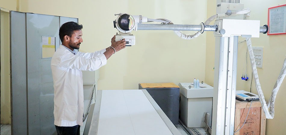
Master of Science in Radiology and Imaging Technology
M.Sc. RIT is a two years course program offered by Ganesh Paramedical College, the best Paramedical College in India located in the country’s capital city Delhi. Master’s program or post graduated programs provided by our college consists of very intensive and comprehensive theory along with regular training facility from the best skilled technologist and high tech farfetched imaging modalities. Given below is the table content of the subjects that are to be covered in the 1st year of M.Sc. RIT.
The details of each subject contents will be found in this blog. Each subject is curated with detailed contents or lessons to be studied and learned in the initial year of this program.
1st year MSc RIT subject code: MSCRIT110
Radiographic Procedure
Theory 100 marks
Unit 1: Basic review of all techniques of Radiography.
Unit 2: Contrast Media related to application, types of contrast, and safety aspects of contrast media/administration mode, administration volume, and techniques of administration.
Unit 3: Anatomy and Physiology of Digestive system, associated pathology and radiographic appearance of digestive system, plain radiography, barium swallow, barium meal, barium meal follow through, enteroclysis, barium enema.
Unit 4: Anatomy and Physiology of Genital and Urinary system, pathology and radiographic appearance of genital urinary system, plain radiography, Intravenous Urography, micturating cystourethrogram, Ascending Urethrography, (ASU), Hystero-salpingography (HSG), Fallopian Tube Recanalization (FTR).
Unit 5: Cardio-Respiratory system anatomy, physiology and associated pathology with radiographic appearance, chest radiography.
Unit 6: Breast anatomy, physiology and pathology; mammography: indications, contraindications and techniques, ICRP guidelines, BIRADS.
Unit 7: Anatomy, Physiology, Pathology and radiographic procedure of the skull overall, facial bones and cranial bones, radiographic projections.
Unit 8: Anatomy, physiology and pathology of the vertebral column and its radiographic projection.
Unit 9: Anatomy, physiology and pathology and radiographic appearances and projection of the upper and lower limbs.
Unit 10: Anatomy, physiology and pathology and radiographic appearances and projection of the Pelvis.
Unit 11: Anatomy, physiology and pathology and radiographic appearances and projection of the Hepatobiliary System, ERCP, PTBD, T-tube cholangiography.
Unit 12: Dental Radiography Anatomy with associated pathology, its radiographic appearances, Intraoral, Extra oral and Occlusal views, General precautions, OPG, mains supply and function of equipment, exposure parameters and radiation protection.
Unit 13: Sialography, Dacrocystography, Sinography, Fistulogram, indications and contraindications of all the mentioned radiographic procedures and their safety measures.
1st year M.Sc. RIT subject code: MSCRIT120
Instrument of Conventional X-ray Equipment
Theory 100 marks
UNIT 1: Generation of electrical energy AC/DC; Polyphase supply Distribution of electrical energy, Use of electrical energy Current loads & power loss, Uses of electricity in Hospitals, Safety rules for Radiographer.
UNIT 2: X-ray Circuit components, high tension transformers, Main Voltage Compensation, High tension switches, Stabilizers and UPS.
UNIT 3: Fuses, Switches, Earthing, High tension cables construction & design. Rectification, Types of Rectifiers, X-ray circuits, Filament circuits, High voltage circuit.
UNIT 4: Tube rating, Types of Generators, Capacitor discharge generator, Battery Powered generator, Battery Powered generator Medium frequency & High frequency generator
UNIT 5: Switches, Circuit breakers, Primary & Secondary switches, Exposure switching and its application. Interlocking Circuits, Regulating and safety devices, Magnetic relay, Thermal relay switches, Interlock in Tube Circuit and overload interlocks.
UNIT 6: Exposure timers, Timing systems: Electronic timer, Ionization timer, Photo timer, Synchronous timer and impulse timer.
UNIT 7: Devices improving radiographic quality/Cone Cylinder/Collimator Grid Filter.
1st year M.Sc. RIT subject code: MSCRIT130
Instrumentation of specialized Radiology Equipment
Theory 100 marks
UNIT 1: Portable & Mobile Equipment, Mains requirements, Cable connections to wall plugs, Portable X-Ray Equipment, Mobile X-Ray Equipment, Capacitor Discharge, Mobile Equipment, Cordless Mobile Equipment, X-Ray Equipment for the Operating Theatre, Mobile Image Intensifier units.
UNIT 2: Fluoroscopy Equipment, Construction & Working principles of Image Intensifier, Viewing the Intensified image, recording the intensified Image, Digital fluoroscopy Panel type image intensifier.
UNIT 3: Fluoroscopic/ Radiographic Tables, General features of fluoroscopic/ radiographic table, the serial changer, Remote control table, spot film devices.
UNIT 4: Tomographic Equipment, Principles of tomography, various types of tomographic movement, Equipment for linear tomography.
UNIT 5: Equipment for Cranial and Dental radiography, the skull table, General Dental X-ray equipment, pantomography equipment, Equipment for Cranial & skeletal radiography, Equipment of mammography.
UNIT 6: Care Maintenance and test, General care, Functional tests, Quality assurance program, Acceptable limits of variation, Corrective action.
1st year M.Sc. RIT subject code: MSCRIT140
Principles of Radiographic Exposure
Theory 100 marks
UNIT 1: X-ray production; Interaction of radiation with matter such as Compton effect, photoelectric effect, pair production, coherent scattering. Useful range Clinical application.
UNIT 2: The Photographic process, Basic review of photographic emulsions, Photographic latent image, Film materials, Spectral sensitivity of film material, Speed and contrast of photographic materials, Intensifying screens and cassettes Film processing.
UNIT 3: Sensitometry, Photographic density, Opacity Transmission, Production of Characteristic curve, Features of Characteristic curve, Variation in the characteristic curve with development, Comparison of emulsions by their characteristic curve, Application of Characteristic curve, Information from the Characteristic curve.
UNIT 4: Radiographic Image Radiographic, Density Acceptable range, Factors influences density. Radiographic Contrast Components, Factors that influence contrast, Management of Radiographic Image quality.
UNIT 5: Resolution, Line spread function & Modulation transfer function, unsharpness in the Radiographic image and various factors contributing towards Unsharpness, Types of Unsharpness, Radiographic mottle.
UNIT 6: Geometry of the radiographic image, Magnification/ Distortion – Types and factors, Micro/ Macro radiography.
UNIT 7: Instrumentation of Processing Equipment Automatic film processor (AFP), Maintenance and Quality control test in AFP, Layout and planning of Darkroom, Viewing accessories: viewing boxes Magnifiers and viewing conditions.
1st year M.Sc. RIT subject code: MSCRIT150
Advanced technique & Instrumentation of Computed Tomography
Theory 100 marks
UNIT 1: Imaging principles in computed tomography, Instrumentation of CT scan, Advances in Detector technology, Slip ring technology, Helical CT, Single slice and Multi slice CT Scan system (recent advancement in CT scanner).
UNIT 2: Isotropic imaging, Image display, Pre and Post Processing techniques, Image quality in single slice and multi slice, helical CT scan, Patient radiation dose considerations in Helical CT.
UNIT 3: Protocols for adult Whole Body CT Protocols for pediatric, Whole Body CT, Documentation, Common and specific artifacts in Helical CT images.
UNIT 4: HRCT of Lungs, Technical aspects, Volumetric HRCT, Expiratory HRCT, HRCT protocols, Artifacts.
UNIT 5: CT angiography, CT fluoroscopy, Multidimensional reformations, MPR, Curved MPT, MIP, 3D imaging & 4D CT.
UNIT 6: CT Perfusion scanning, Dentascan, CT colonoscopy, CT bronchoscopy.
UNIT 7: CT coronary angiography, CT calcium scoring, Myocardial Imaging.
UNIT 8: Care, Maintenance and tests, General care, Functional tests, Quality assurance program, Acceptable limits of variation, Corrective action.
You would be able to locate Ganesh Paramedical College in Rohini, to start off your journey to a Paramedical Expert. The number of seats are Limited to we expect the students to contact as soon as possible to grab and secure this opportunity. So, join us in this healthcare ride of success. Hurry up! Contact Now!