Exploring the Advancement of X-Ray Modality in Medical Province

Updated Post: 04 May 2024
Share Post:
![]()
![]()
![]()
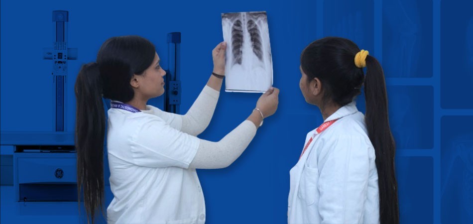
Radiography was introduced more than 100 years ago by German scientist Sir Wilhelm Conrad Roentgen in 1895. The first Nobel Prize to be awarded for Physics in discovery of X-ray was presented to Sir Roentgen in the year 1901. In today’s time just as all other fields across varieties of industries, digital advancement is transmogrifying the field of radiography.
A lot of advancement has been made and is still being made in the field of Digital radiography including AI aided interpretation of X-ray, DE imaging or Dual-energy imaging, diagnosis that are computer-aided, tomosynthesis, digital mobile radiography, and automatic image stitching.
The advancements lead to improved quality of image with enhancement towards patient care and better patient outcomes. Additionally, digital radiography decreases retake of exposures leading to low radiation exposure benefits.
Advantages of Digital Radiography
- Less exposure time
- Improved detail evaluation
- High image quality
- Fast image processing
- Clearer diagnosis
- Easy data and image storage
- Proficiency for remote viewing
Few comparisons between conventional or old ways of x-rays and modern digital x-rays are given below:
Capturing of Image: Old style x-ray system consisted of usage of films and cassette sandwich where images were captured by exposing the radiation that produced a latent image on the film. Latent image is also known as raw data. In a positive and advanced comparison, digital radiography use electronic detectors to capture images where the x-ray photons are converted into digital data to be viewed on a computer monitor.
Comparison of Image Quality: In conventional x-rays, list of factors goes into image quality such as the film quality condition, exposure techniques and processing standards. Additionally, several attempts may be required to achieve the desired image quality. Higher image quality and precision are offered easily by digital x-ray modalities provided with the capability of the digital images to be enhanced, manipulated and edited to improve visualization that can aid to a more accurate evaluation of anomalies. These digital images can be viewed and analyzed instantly that allow on the spot adjustment of the image quality if necessary.
Radiation Exposure: In the old style x-rays system, higher radiation doses were required to give out adequate quality images which again sometimes had to be repeated to achieve a good image quality for diagnosis. Digital x-ray systems utilize lower radiation dose and also maintain image quality at the same time because the exposure settings can be adjusted to an accurate and optimum precision. These functions ultimately reduce radiation exposure to patients as well as healthcare workers.
Operation and Efficiency of Work: Manual processing of films was done under conventional x-rays which was quiet time-consuming and includes physical labor. The films are first developed then fixed and then manually inspected before evaluation. In digital modality for x-rays, the images are immediately available and presented right after exposure with the complete elimination of manual labor for image processing resulting in increased efficiency and a properly streamlined workflow. The resultant data can be easily stored, sent, and accessed easily which helps greatly in faster evaluation of pathology and planning of treatment.
Physical storage space and PACS: Conventional X-ray films need a physical storage room or some sort of space with very careful maintenance to prevent any kind of damage or degradation as time passes. To retrieve a specific image is time-consuming as it requires manual searching. Picture Archiving and Communication System refers to digital storage systems that allow image data storage in an electronic manner that allows easy storage, access to retrieval anytime effortlessly, and sharing of images among healthcare facilities, ultimately leading to saving time and improved patient care.
Conclusion
Modern Day X-ray has advanced leaps and bounds and so has the education system at Ganesh Paramedical College. We make sure to be updated with our teachings and impart quality education to our students. To ensure that our students get 100% assistance in jobs and the best minds get placed at Ganesh Diagnostic and Imaging Centre itself. So, enrol yourself at the best paramedical college in India, Ganesh Paramedical to secure your future by learning on the high tech machines and latest and advanced technologies. For seeking admission with limited seats at our prestigious college, you are requested to contact us on a first-cum first basis.
Connect to us at any of the platform given below! Hurry up! Enrol now!